Me too! Well until a couple weeks ago I would have had to take some shrooms and imagine up some images of gold atoms. Not anymore.

Where these two gold crystals meet they are joined by a complex arrangement of atoms, forming a nanobridge that accommodates their different orientations. The atoms are 2.3 angstroms apart. TEAM 0.5's unprecedented signal-to-noise ratio makes it possible to distinguish individual atoms and, at the edges of the two crystals, deduce their position in three dimensions.
Thanks Science! What wonderful machine done brought us these fine images? This beast.
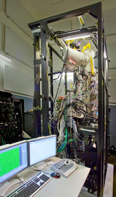
TEAM 0.5, the world’s most powerful transmission electron microscope — capable of producing images with half-angstrom resolution (half a ten-billionth of a meter), less than the diameter of a single hydrogen atom also known as really freaking small — has been installed at the Department of Energy’s National Center for Electron Microscopy (NCEM) at Lawrence Berkeley National Laboratory.
These guys don’t just look at stuff that is small, they run the operation like the it is some sort of battle ship, or space ship. “We have beam down the column,” announced Uli Dahmen of Berkeley Lab’s Materials Sciences Division, when the TEAM 0.5 microscope first delivered its ultrabright electron beam at Berkeley Lab in late December. How cool would that be to be able to scream things like “WE HAVE BEAM!!!” I love science.
The TEAM Project (TEAM stands for Transmission Electron Aberration-corrected Microscope) is led by Berkeley Lab in a collaboration with DOE’s Argonne and Oak Ridge National Laboratories, the Frederick Seitz Materials Laboratory of the University of Illinois, and two private companies specializing in electron microscopy, the FEI Company headquartered in Portland, Oregon, and CEOS of Heidelberg, Germany.
Now that TEAM 0.5’s basic systems are operational, additional components and facilities are being completed and tuned, including a state-of-the-art control room display that shows the sample under the microscope on a flat panel resembling a wide-screen, high-definition TV. After a long series of rigorous tests and adjustments, TEAM 0.5 will become available to outside users by October, 2008.
Atom by atom in 3-D
In preliminary tests at the FEI Company, before the TEAM 0.5 was shipped, NCEM’s Christian Kisielowski tested the microscope’s ability to resolve individual atoms and precisely locate their positions in three dimensions. He made a series of images of two gold crystals connected by a “nanobridge” only a few dozen atoms wide. From each exposure to the next, individual gold atoms could be seen changing positions.
He watched atoms wiggle, its like watching your DNA wiggle. There is something so profound about being able to SEE atoms wiggle. I would feel like some sort of super being if I got to do that. “Hey what did you do today?” pause “Oh I ran som errands, then spent the rest of the day staring raptly at the building blocks of matter!!!”
To achieve this extraordinary resolution, TEAM 0.5 embodies technical advances that have only recently become possible, including ultra-stable electronics, improved aberration correctors, and an extremely bright electron source. In short, they are the new techno-gods of looking at small things.
Spherical aberration degrades images, making points of light look like disks, and correcting it can make dramatic improvements to image resolution. (This was famously demonstrated in 1993, when spherical aberration in the Hubble Space Telescope’s optical lenses was corrected in a special space mission.) In the case of electron microscopes, a series of multipole magnetic lenses of varying geometries shapes the electron beam.
“Correcting spherical aberration in an electron microscope has long been possible in theory,” says Dahmen. “But only recently has it become practical,” because today’s stable electronics reduce drift and fast computers allow continuous adjustments in real time. Corrector technology has even become available commercially, says Dahmen, “but no off-the-shelf corrector can match TEAM 0.5’s ability to compensate even higher-order aberrations.”
Correcting spherical aberration makes it possible to use the TEAM 0.5 not only for broad-beam, “wide-angle” images but also for scanning transmission electron microscopy (STEM), in which the tightly focused electron beam is moved across the sample as a probe, capable of performing spectroscopy on one atom at a time — an ideal way to precisely locate impurities in an otherwise homogeneous sample, such as individual dopant atoms in a semiconductor material.
Aberration correction is also essential for another advanced feature of TEAM 0.5: its ability to maintain high resolution with lower electron beam energies.
“Low-energy electrons have longer wavelengths, so they are harder to focus,” Dahmen explains. “Aberration correction allows better than one-angstrom resolution with excellent contrast even at 80 kilovolts. This is important when you don’t want to damage the sample with a high-energy beam — in biological studies, for example.”
It’s not just high resolution that makes TEAM 0.5 the world’s best microscope, Dahmen says. When all the electrons in the beam focus at the same plane, image contrast and signal-to-noise ratio improve tremendously.
“It’s because the signal-to-noise ratio is so good that you can adjust focus atom by atom, with enough sensitivity to obtain information about the three-dimensional atomic structure of a single nanoparticle.” Dahmen adds, “This brings us within reach of meeting the great challenge posed by the famous physicist Richard Feynman in 1959: the ability to analyze any chemical substance simply by looking to see where the atoms are.”
I am sorry to geek out here, but HOLY CRAP, like you know you look at a tree and you go “ohh thats an oak tree” cause you know what oak trees look like, imagine if you could look at something and go “ohh thats hydrochloric acid” cause you know what hydrochloric acid looks like at an atomic level.
The position of individual atoms in a structure can be determined by taking images at different angles, from which the computer reconstructs a 3-D tomograph of the sample, as in a CAT scan. To make this possible an innovative system capable of tilting and rotating the sample, and moving it up, down, or sideways under the electron beam, is also being developed at NCEM.
Much smaller than sample stages now in use, the new TEAM stage will be housed entirely inside the microscope column. Manipulating the sample by such methods as minute piezoelectric “crawlers” that change shape when electricity is applied, the new stage will be able to control and reproduce the sample’s position and attitude with an accuracy of less than a billionth of a meter.
Installation of the new stage must await the next phase of the TEAM Project: the TEAM I microscope, due to be set up at NCEM early in 2009.
While TEAM 0.5 corrects spherical aberration in both the “probe” beam (the electron beam before it strikes the sample) and the image beam (after it exits the sample, but before it reaches the detector), TEAM I will also correct chromatic aberration in the image beam, which has never been accomplished before. Spherical aberration is caused by the shape of a lens; chromatic aberration results when a lens refracts light or electrons of different wavelengths (different colors or energies) at different angles.
“Correcting chromatic aberration is harder and takes more space,” says Dahmen. “The chromatic aberration corrector will add two feet to the height of the TEAM I column. But the new configuration will also allow us to enlarge the gap between the pole pieces, into which the sample fits. In TEAM 0.5 this gap is only about two millimeters, so we have to use traditional outside-mounted sample stages, with limited space to manipulate the sample. In TEAM I the gap will be five millimeters; the sample stage will have much greater freedom of movement.”
New vistas in the realm of the small
TEAM 0.5 and TEAM I will be housed side by side at NCEM for some time, occupying the two multistory “silos” that until recently were the homes of the historic High-Voltage Electron Microscope and the Atomic Resolution Microscope, the most powerful microscopes in the world when NCEM was established in the early 1980s.
Ambitious as those microscopes were in their day, says TEAM’s Project Manager, Peter Denes of the Engineering Division, “when the TEAM Project was launched in 2004, it was not quite clear if the goals could even be achieved. The electron microscopy community had never done a collaborative project like TEAM before, and certainly not with full DOE project-management rigor.”
Says Denes, “Perhaps the biggest contributor to success was a series of scientific workshops that contributed to forming a converging opinion on what the next steps would be, and what would constitute success. That helped in getting everyone — if not quite on the same page — at least in the same book.”
Dahmen agrees. “This is a big jump for the microscopy community. TEAM’s success will open the door to other ambitious developments around the world.”
Dahmen suggests at least two broad categories of researchers who will benefit from the powerful new electron microscopes: experts with sophisticated microscopy problems to solve, and scientists less familiar with electron microscopy but with a particular problem for which microscopy can provide the answer.
“For example, Jim Zuo at the University of Illinois is doing studies of electron diffraction from the surface of single nanoparticles,” Dahmen says. “He sees evidence of surface contraction. But when we at NCEM do imaging of similar nanoparticles, we find that the surface is expanding. Jim looks forward to using the TEAM microscope because it can do diffraction and imaging of the same particle at the same time — a grand experiment, and the only way to solve the apparent contradiction.”
An example of a problem-solving nonspecialist, says Dahmen, might be a materials scientist who has created a new kind of nanostructure, such as a tetrapod semiconductor, and needs to know exactly where in this complex, three-dimensional shape the impurity atoms reside. “TEAM’s ability to image the structure in 3-D through tomography and its ability to do spectroscopy with single-atom sensitivity can identify each kind of atom at each position in the structure. That has never been possible before.”
The basic TEAM components of aberration correction, enhanced signal-to-noise ratio, single-atom sensitivity, and an ultrabright beam that can be used in both TEM and STEM modes — all the while manipulating the sample in the beam — are goals that until recently seemed at the very edge of technological daring. All are on track, and some have been solved ahead of schedule. The TEAM Project’s continuing success, signaled by the installation of TEAM 0.5 at NCEM, has opened the possibility of numerous future advances in electron microscopy that were barely conceivable when TEAM was launched.
I can’t wait to see what they come up with next.
Wow this is nice
At first, upon close inspection the ‘grain boundary’ region in the centre appeared to contain an additional two single crystal orientation bands approximately 7 atoms thick. But then I saw the interface was actually an ‘s-shape’ with 120 deg interior angle, with that boundary atoms themselves exhibiting intermediate orientation respective of their adjoining single crystals. Fascinating to actually ‘see’ such a theoretical structure so clearly.
I think this image contains a lot of info. It looks like some sort of cubic structure just from the unit pattern. It would be really neat to be able to see some interior, non-edge dislocations as well.
why would we wanna see gold atoms …???
whats the use … ???
gold is very stable and has a nice crystal structure, so I guess thats is why they went with gold…but they could have done this with just about anything.
I don’t get what I am seeing here. Why can’t we see atoms on the left & right sides ( but only in the center)? What would this look like under the naked eye, just 2 gold crystals touching?
I didn’t even know gold could crystallize.
Back to school for me I guess. :(
What you are looking at is two very very skinny pieces of gold that are touching each other. The reason why you don’t see any on the left and right is because that is empty space (or mostly empty space).
Many metals, and many other elements are “crystals” that just means that the atoms inside the material line up in a repeating pattern. (this is the highly simple explanation)
Why do the atoms look so ‘big’ compared to the space between them? I mean, I thought most of of an atom was just empty space, with the nucleus being a very, very tiny part of the space an entire atom takes up. Why does the nucleus look large? Shouldn’t there be more empty space between them?
my understanding of it is that them dots you see are the nuclei AND the electron cloud…so the dot is the whole works, and thus the dot itself is mostly empty space. Fascinating how much of what we consider “solid” is just empty space.
So how far from this to a molecular assembler? Is there any scientific problem with nanotechnology going this far or is it just an engineering problem?
this just looks at stuff, moving atoms around, and the like I think would take different technology
You can see the same thing with Scanning Tunneling Microscopy, you don’t need such a thin piece of gold either. Granted, you get a topographical image from STM, but it’s a more passive technique. Google “STM images” and you can see all kinds of molecular and atomic resolution stuff. Don’t get me wrong, this is a great technique; I did some TEM today.
very nice to read
This is such a great post! Not only is the subject matter really freaking cool, but it’s good to see some one else geeking out about things like this. Keep it up!
cool Atom!!!!!!!!!!!!!!!!!
cool thing it looks realllllly cooool!!!!!!!!!
gold is my favorit!!!!!
gosh..are they real atoms.. cant believe..but how can the atoms are supposed to be circular?? i mean from wat we have learnt that atoms dont have boundaries…they are like mere shadows isnt it…but anyway the pic’s a real beauty
I wear a lot of gold
hey people that is reading diz……. sooooo what yall wanna talk about..well lets talk about gold… yes jajajaja.. anyways
hey people that is reading diz……. sooooo what yall wanna talk about..well lets talk about gold… yes jajajaja.. anyways… talk to yall later
hey wat up wat you all doing
gold is goooooooooood
i want to know wat propities gold has in it, could you put it in?
gold balls
“gosh..are they real atoms.. cant believe..but how can the atoms are supposed to be circular?? i mean from wat we have learnt that atoms dont have boundaries…they are like mere shadows isnt it…but anyway the pic’s a real beauty”
although it looks good, it is not really a picture of atoms. A scanning electron microscope dose not allow you to SEE things the way a regular microscope dose. Instead it passes a charged probe across the surface of an object and measures slight changes in electricity flowing through the probe. It then sends that data to a computer, which uses a complicated set of mathematical equations from a theory called “quantum mechanics” to calculate what she surface of the object should look like. The computer then graphs the results of that calculation, adding colors to enhance the quality of the image.
so what you are really seeing in this photo is the result of a calculation that comes from a theory about how electricity flows under certain circumstances. Thus if the theory is correct, and if the computer calculation is correct, then you are seeing a REPRESENTATION of atoms on the surface of the metal examined with the scanning electron microscope. Those are two big “if’s” however.
Now I personally think both the theory and calculations are correct, so I think that what you see in the image above is probably a good representation of gold, excluding the color, which has been artificialy added.
Reply to mad octopus’ “are they real atoms?” question.
Although it looks good, it is not really a picture of atoms. A scanning electron microscope dose not allow you to SEE things the way a regular microscope dose. Instead it passes a charged probe across the surface of an object and measures slight changes in electricity flowing through the probe. It then sends that data to a computer, which uses a complicated set of mathematical equations from a theory called “quantum mechanics” to calculate what she surface of the object should look like. The computer then graphs the results of that calculation, adding colors to enhance the quality of the image.
so what you are really seeing in this photo is the result of a calculation that comes from a theory about how electricity flows under certain circumstances. Thus if the theory is correct, and if the computer calculation is correct, then you are seeing a REPRESENTATION of atoms on the surface of the metal examined with the scanning electron microscope. Those are two big “if’s” however.
Now I personally think both the theory and calculations are correct, so I think that what you see in the image above is probably a good representation of gold, excluding the color, which has been artificialy added.
This looks like what we would conventionally consider atoms to look like. Round circular objects. But how do you know it’s not just another representation of a crystalline structure?
And even if they say quantum tunneling gathers information on the scale of an electron, shouldn’t an accurate picture of the atom show atomic structure, which is not present?
Hey, so yeah you guys should you now realy show the whole atom, I mean yeah I now you guys are doing your best but I mean c’mon do better!
You people are dumb! No whole atom, no money!
GOLD REALY! MEST-UPPPPPPPPPPPPPPPPPPPPPPPPPPPPPPPPPPPPPPPPPPPPPPPPPPPPPPPPPPPPPPPPPPPPPPPPPPPPPPPPPPPPPPPPPPPPPPPPPPPPPPPPPPPPPPPPPPPPPPPPPPPPPPPPPP!!!!!!!!!!!!!!!!!!!!!!!!!!!!!!!!!!!!!!!!!!!!!!!!!!!!!!!!!!!!!!!!!!!!!!!!!!!!!!!!!!!!!!!!!!!!!!!!!!!!!!!!!!!!!!!!!!!!!!!!!!!!!!!!!!!!!!!!!!!!!!!!!!!!!!!!!!!!!
YOUR RIGHT JADEN MEST-UPPPPPPPPPPPPPPPPPPPPPPPPPPPPPPPPPPPPPPPPPPPPPPPPPPPPPPPPPPPPPPPPPPPPPPPPPPPPPPPPPPPPPPPPPPPPPPPPPPPPPPPPPPPPPPPPPPPPPPPPPPPPPPPPPPPPPPPPPPPPPPPPPPPPPPPPPPPPPPPPPPPPPPPPPPPPPPPPPPPPPPPPPPPPPPPPPPPPPPPPPPPPPPPPPPPPPPPPPPPPPPPPPPPPPPPPPPPPPPPPPPPPPPPPPPPPPPPPPPPPPPPPPPPPPPPPPPPPPPPPPPPPPPPPPPPPPPPPPPPPPPPPPPPPPPPPPPPPPPPPPPPPPPPPPPPPPPPPPPPPPPPPPPPPPPPPPPPPPPPPPPPPPPPPPPPPPPPPPPPPPPPPPPPPPPPPPPPPPPPPPPPPPPPPPPPPPPPPPPPPPPPPPPPPPPPPPPPPPPPP!!!!!!!!!!!!!!!!!!!!!!!!!!!!!!!!!!!!!!!!!!!!!!!!!!!!!!!!!!!!!!!!!!!!!!!!!!!!!!!!!!!!!!!!!!!!!!!!!!!!!!!!!!!!!!!!!!!!!!!!!!!!!!!!!!!!!!!!!!!!!!!!!!!!!!!!!!!!!!!!!!!!!!!!!!!!!!!!!!!!!!!!!!!!!!!!!!!!!!!!!!!!!!!!!!!!!!!!!!!!!!!!!!!!!!!!!!!!!!!!!!!!!!!!!!!!!!!!!!!!!!!!!!!!!!!!!!!!!!!!!!!!!!!!!!!!!!!!!!!!!!!!!!!!!!!!!!!!!!!!!!!!!!!!!!!!!!!!!!!!!!!!!!!!!!!!!!!!!!!!!!!!!!!!!!!!!!!!!!!!!!!!!!!!!!!!!!!!!!!!!!!!!!!!!!!!!!!!!!!!!!!!!!!!!!!!!!!!!!!!!!!!!!!!!!!!!!!!!!!!!!!!!!!!!!!!!!!!!!!!!!!!!!!!!!!!!!!!!!!!!!!!!!!!!!!!!!!!!!!!!!!!!!!!!!!!!!!!!!!!!!!!!!!!!!!!!!!!!!!!!!!!!!!!!!!!!!!!!!!!!!!!!!!!!!!!!!!!!!!!!!!!!!!!!!!!!!!!!!!!!!!!!!!!!!!!!!!!!!!!!!!!!!!!!!!!!!!!!!!!!!!!!!!!!!!!!!!!!!!!!!!!!!!!!!!!!!!!!!!!!!!!!!!!!!!!!!!!!!!!!!!!!!!!!!!!!!!!!!!
Fuck!
happy new year!!!
The response level to national disaster is noble but it’s a real shame that so many people take advantage of the sad situations.
I mean everytime there is an earthquake, a flood, an oil spill – there’s always a group of heartless people who rip off tax payers.
This is in response to reading that 4 of Oprah Winfreys “angels” got busted ripping off the system. Shame on them!
http://www.cbsnews.com/blogs/2009/08/19/crimesider/entry5251471.shtml